
 Tiếng Việt
Shrimp get sick too. Taura syndrome is an infectious disease caused by the virus TSV (Taura syndrome virus). TSV infects tissues of ectodermal and mesodermal origin including whole body hypodermis (cuticular epithelium) and gills. However, endoderm-derived organs such as the hepatopancreas, midgut, midgut caeca, muscle and heart don´t show histological evidence of TSV.
Taura syndrome was first reported in P. vannamei from the Taura region in Ecuador in 1997 and then promptly spread to other American countries and other continents including Asia. Ecuadorian shrimp farmers first believed the disease was caused through the use of pesticides in banana plantations close to shrimp farms, however, scientific research confirmed it’s a viral etiology. TSV is a reportable OIE disease.
It is suspected that TSV causes increased health problems in densely populated aquatic animal production environments. Taura syndrome disease may occur more often when salinity is below 30 ppt. Four different TSV genotypes have been reported: group 1: Americas, group 2: South-East Asia, group 3: Belize, and group 4: Venezuelan, each having different commercial impacts.
TSV infection is characterized by mass mortality (40% – 90%) and is observed at the early stage of juvenile farming 14-days after pond stocking. TSV can spread between pond populations through horizontal transmission (cannibalism) and virus detection in early post larvae suggests that vertical transmission is also possible. TSV infection can be detected by PCR analysis in almost all shrimp life stages including post larvae, juveniles and adults of P. vannamei but not in eggs, larvae or zygotes.
Causative agents of Taura syndrome. The pathogenic agent TSV belongs to the Genus Aparavirus, Family Dicistroviridae, Order Picornavirales. TSV can be transmitted to susceptible shrimp populations by vectors that include infected feces of Sea Gulls (Larus atricilla), chicken (Gallus gallus) and aquatic insects like water boatman (Trichocorixa reticulate).
Clinical signs of TSV. The appearance of clinical signs can occur as early as late post larvae or juvenile stages in nursery or growout ponds. Three disease phases have been differentiated.
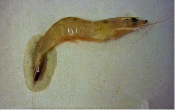
Acute phase (pictured right): Shrimp in terminal decline display red chromatophores (pigmented cells) expansion, general pale reddish discoloration, distinctly red pleopods, soft shell, empty gut and are often sick in the late D stage of the moult cycle (post-moult). Severely affected shrimp die during C-D stage of molting (ecdysis). Only during the acute phase is it possible to observe pathognomonic (disease specific) histological findings characterized by multifocal necrosis in cuticular epithelium of the body surface, gills, appendages, hindgut, esophagus, and anterior and posterior stomach chambers. The antennal gland can also be destroyed in severely affected organisms due to infection.
In addition to histological diagnosis, examination under light microscope wet mounts can be useful to demonstrate TSV in sick shrimp during the acute phase by observing abundant spherical dark structures (necrotic cells), characterized by presence of pyknotic (condensed) and karyorrhectic (fragmented) nuclei and cytoplasmic remnants.
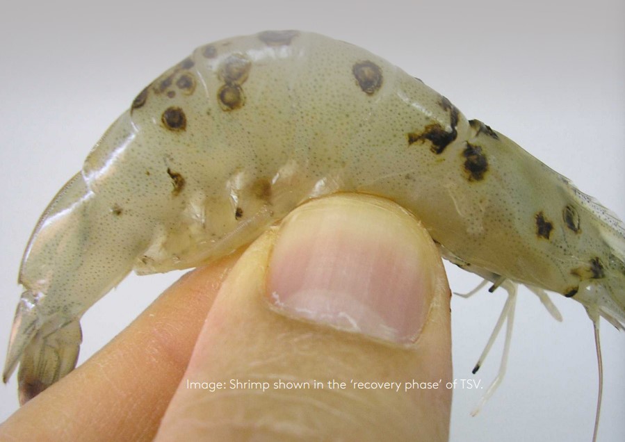
Recovery phase (pictured front cover): Shrimp that survive the acute phase then enter a recovery phase. Common clinical signs during the recovery phase include dark–black or dark-brown randomly placed multifocal and irregularly shaped cuticular melanized lesions. These are sites of resolving TSV lesions in the cuticular epithelium. Sick animals in this phase feed normally and may have soft shells and generalized expansion of red chromatophores.
Chronic phase: When shrimp recuperate from recovery phase and cuticular melanized lesions disappear via moulting, they establish a new healthy exoskeleton with no evidence of TSV disease. These ‘survivor’ shrimp may remain infected throughout their life with no clinical signs, however they are less resistant to environmental stressors in the intensive farming environment.
Early detection of TSV using Shrimp MultiPath™. Early testing and detection with Shrimp MultiPath™ can give farmers two to six week’s notice before clinical signs appear and prior to mass mortalities. In commercial nursery ponds and growout shrimp ponds, TSV infection can be detected early, and farmers advised as soon as juveniles are stocked in ponds. This information is an early warning system preparing farmers for a critical period where slowing the spread of the disease and maximizing production outputs is still possible.
Early detection empowers the implementation of prompt mitigation strategies. These can include:
- PCR assays for pre-screening of broodstock before placing in production tanks
- PCR assays for pre-screening of post larval shrimp discarding tanks that test positive for TSV infection
- Suspending pond stocking with post larvae from infected hatcheries
- Avoiding live and fresh feeds (especially for broodstock) from countries with historic status of TSV infections
- Not feeding female broodstock 6 hours before moving to spawning tanks to reduce egg contamination with feces, and reinforcing eggs and nauplii washing and disinfection before transferring to hatchery tanks to reduce possible TSV contamination from broodstock feces
- Use post larvae from breeding programs focused on exclusion plans and production of TSV-Free or Specific Pathogen Resistant or Free (SPR/SPF)
- TSV-resistant or tolerant post larvae pond stocking only with TSV PCR tested post larvae and, pond surveillance for TSV using molecular tools.
Preventive farming strategies that may reduce TSV transmission include:
- Restocking of entire farming zones with TSV-free stocks
- Removing sick or dead shrimp to prevent transmission through cannibalism
- Reducing pond density (partial harvest)
- Organic debris and feces removal (syphoning and/or bacterial bioremediation when possible),
- Proper technical assistance for periodic monitoring with appropriate diagnostic tools will allow for discrimination between TSV and other disease with similar clinical signs
- Biosecurity around infected ponds must be increased, for example management of affected ponds in daily routines, separating nets and equipment, physical barriers put in place, inform adjacent farmers of the infection, and harvest when commercial size is reached. Disease mitigation plans should include pathogen exclusion programs.
Reference: https://genics.com.au/wp-content/uploads/2021/10/Taura-Syndrome-Virus-TSV-Guide.pdf?x60024
DOMESTICATED SHRIMP POSTLARVAE – THE KEY TO SUCCESS
See more:
- Heat wave causes a huge loss to the Satkhira shrimp farmers
- Can dietary inclusion of montmorillonite clay help mitigate the farmed shrimp AHPND pandemic?
- Scientists suggest krill meal is key to obtain cost-effective and nutrient-rich shrimp feed formulations